鈴鹿・四日市・亀山で、歯科・矯正歯科・インプラント・ホワイトニング・各種保険診療のことなら大木歯科医院
 鈴鹿インプラント矯正クリニック
三重県鈴鹿市南長太町鎗添2504-2 受付時間 8:30より 診療時間 9:00〜19:30

 059-395-1000
 ohkident@mecha.ne.jp
AAIDとは??? http://www.aaid-implant.org
1951年に設立された、アメリカでもっとも歴史の長い学会です。
古くはLINKOW先生、新しいところでは、CARL MISCH先生など、アメリカのインプラント学を引っ張ってきた学会です。もうひとつ、有名なのが、AO(Academy of Osseointegration)ですが、これはもともとブロンネンマルク教授学派によって1982年(HPより)に設立された新しい学会です。資格社会である米国において、現在AAIDとABOI(アメリカンボード;但し、これは米国またはカナダの歯科医師免許が必須)がアメリカ社会においてインプラント治療についての広告が許可されています。これは、AAIDの300時間にわたる講義とその後に行われる記述試験/症例試験/口頭試問など厳正な資格試験が米国社会で認められた結果です。(厳密には、各州の裁判所でそれが審議され、米国の主だった州で、AAIDのインプラント専門医認定資格を広告することが認められています。)
という内容を愛知インプラントセンターの堀田先生からご紹介頂きました。
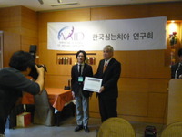
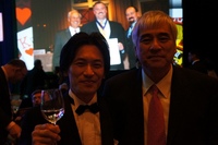 アメリカ口腔インプラント学会授賞式にて堀田先生と記念写真
「海外に出なさい」という寺西先生の言葉がどこかに引っかかっていました。
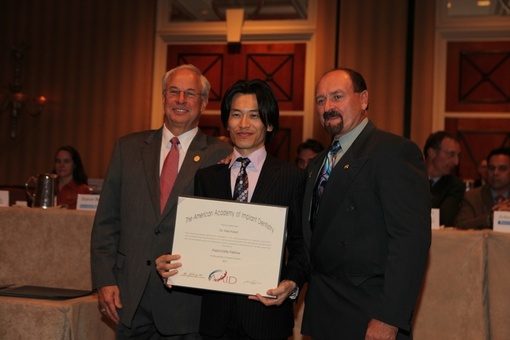
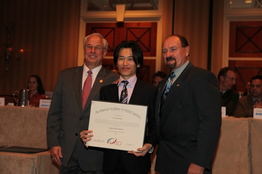
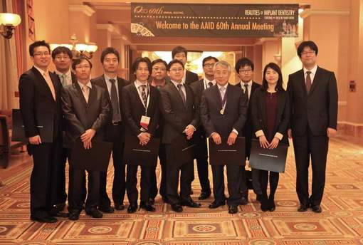
Dr.Norman 彼は60年前に第1回アメリカ口腔インプラント学会を開催しました。
という事はアメリカには60年前、すなわち1951年にはインプラントが行われ始められていたという事です。
日本でインプラント治療が社会的に認知され始めたのはこの20年くらいですが、それよりも遥か昔からアメリカではインプラント治療が行われており、成功と失敗の経験が蓄積されているのです。
インプラント治療の成功率を飛躍的に向上させる為にはインプラント治療の歴史から学ぶ事が不可欠と考えました。さらには最新の技術を学ぶ事が出来ることにも魅力を感じました。
最新技術についてはすぐに患者様に導入するのではなく、日本よりも先駆けて行われている臨床データを見届けた後、実際に院内に導入するというスタンスが大木歯科医院の方針です。
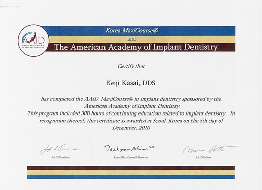
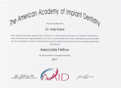
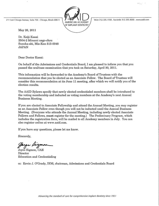
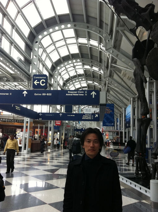
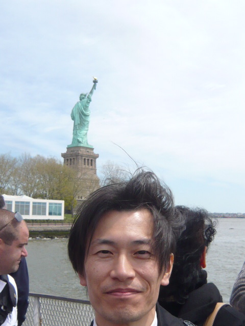
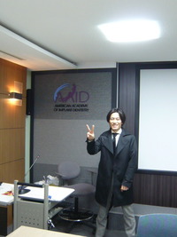
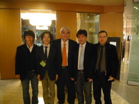
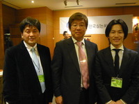
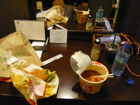
アメリカ口腔インプラント学会AAIDの認定取得のための300時間のセミナーが終わると1回目の試験が待っています。往復の飛行機の中とホテルでは試験勉強。食料を買い込んで周囲の観光客の方が楽しそうにしていても我々は部屋にこもって受験勉強。やり残した仕事を持ち歩きながら、待ち受けている試験のプレッシャーで焦るばかり。気持ちはいつも受験生、、、でした。
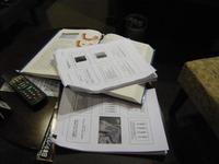 試験の資料 山積み!
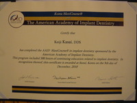 300時間セミナー受講証

AMERICAN ACADEMY OF IMPLANT DENTISTRY
211 East Chicago Ave, Chicago IL 60611-2616 312/335-1550
CHECKLIST FOR ELECTRONIC CASE REPORTS FOR THE ASSOCIATE FELLOW EXAMINATION
Instructions: Place an "x" before each item that is included in the case report. To verify that you have personally reviewed this report and checklist for accuracy, write your Examination Number in the space provided at the end of the checklist. Include this checklist in each of your written case reports as the first page.
This case meets the following case requirement for Associate Fellowship:
x Single Tooth
Edentulous Segment of Two or More Adjacent Teeth with a minimum of two implants
Completely Edentulous Arch
This case report includes the following as specified in theGuidelines for Case Reports for Associate Fellow Membership andInstructions for Submission of Electronic Case Reports:
A narrative (prose) report
My report includes the following sections
Patient Examination
Development of the Treatment Plan
Surgical and Prosthetic Report
Clinical Resume
A health history with the patient's signature
Treatment consent form with the patient’s signature
The required radiographs
Each radiograph is labeled as specified
The required post-completion photographs
Each photograph is labeled as specified
A signed patient release form for the case is submitted with my case report
With the exception of the patient release form, myname, office name, and office address do not
appear anywhere in this case report.
My candidate number is AF1182
Candidate number: AF1182
Patient: AW
Case Type:
Medical history |
|
Insert a scanned copy medical history with patient’s signature. Click on the sample history below, go to INSERT picture and choose a scanned copy of the medical history. BE SURE SCAN is LEGIBLE.
|
|
|
![]()
![]()
![]()

![]()
|
|

|
|
![]()
![]()
|
|
||||||||||||||||||
|
|
|||||||||||||||||||
|
|
|
|
|||||||||||||||||
|
|||||||||||||||||||
|
|
|||||||||||||||||||
|
|
|||||||||||||||||||
|
|
|
|
|||||||||||||||||
![]()
![]() nurse
nurse
|
How did you do it? |
□I have a pain in a tooth(A tooth soaks、A throb is painful、I have a pain in it when I get caught、I swelled up、Others) □ There is bad breath □ Bleeding from the gums □There is a blotch in a mouth □ A denture does not fit/I want you to make a denture □A face swells up ■I want to take the tartar □ I want you to check-up it ■Others( ) |
|
How did you know this doctor's office? |
□The introduction from an acquaintance / a family(The introducer name: ) □ To see a station signboard □ To see a building □ It is a family dental clinic □To see a telephone book □ To see a homepage □ Because it is near □Others( ) |
|
It having been taken dental treatment last time? |
□in this office ( month before) □in other office ( month before) |
|
The impression at that time? |
□ I had a pain in it □ I did not have a pain in it □ I was fierce □ I was tender □ Going to hospital was difficult □ It was convenient ■I was not able to receive enough explanation □I was able to understand well □others( ) |
|
If there is hope about medical examination and treatment |
□ I want you to finish treatment by ( ) □ I cure a lot at a time and I want to reduce the number of times of the going to hospital as much as possible □ Even if the number of times of the going to hospital increases, I want you to do one treatment a little □ I am extremely easily scared □ I want you to cure only a place having a pain in, a troubled place
■ I want you to cure of a place having a pain in, problems in not only a troubled place but also all oral problems □ I want you to arrange the time for counseling (consultation) about my tooth |
|
If there is the hope about the reservation |
・The time when I can visit a hospital for treatment□AM □PM ・ The day when I can visit a hospital for treatment ・ □mon □tue □wed □thu □fri □sat |
|
The current state of health |
■good □normal □bad ※Only as for the woman □The pregnancy( month) |
|
The disease that appeared so far |
■n.p. □Diabetes□cardiac disease □High blood pressure □Low blood pressure □Kidney disease □A liver disease □Tuberculosis □Hepatitis □others( ) |
|
Is there a thing such as the next? |
□A wound is easy to fester □Urticaria is easy to appear □A rash is easy to come out □It is easy to suffer from stomatitis □Medicine hypersensitivity (a medicine name: ) □It is easy to have loose bowels □There is asthma □It is easy to catch cold □Blood is hard to stop □A stomach worsens when I take medicine □It is pollinosis □Others( ) |
|
Is there the medicine taking now? |
■nothing□A vitamin compound□A hormone drug □An antihypertensive agent□A medicine for diabetes□others( ) |
|
Have I pulled a tooth before? |
□no ■yes ⇒Was there the abnormality then? □I came to feel sick □Blood was hard to stop □others( ) |
|
Blood pressure |
□high ■normal □low Maximum: Minimum: |
|
In addition, if there are becoming going and hope, please fill it out |
|
Patient Examination |
|||
History[Describe the chief complaint and secondary complaints. Be sure to describe the patient’s medical history as well as any laboratory findings (e.g. CBC, SMA, PTT, INR) and current medications, as applicable.] I want you to remake a bridge at 567 parts of lower right. Dirts are easy to collect around pontic part and are hard to clean. I am worried about the metal color of the bridge. In addition, there is no systemic anamnesis Important Notice.
|
|||
Clinical Examination[Describe the existing dentition, adjacent soft tissues, periodontal charting, lip line, temporomandibular joint function, parafunctional habits, hard and soft tissue anatomy of edentulous areas and other findings.] The existing dentition 76543211234567 75 43211234567
Iip line:Normal Oral hygiene:It is not slightly good Periodontal problem:Slight gingivitis Temporomandibular joint function:n.p. Parafunctional habits(-) The volume of bone is enough vertically and horizontally. There is enough volume of attached keratinized gingival at buccal side. Parafunction(-).
|
|||
Radiographic Examination[Describe the findings and limitations.] I was able to confirm the healing of socket in a preoperative panorama photograph. Distance to a mandibular canal is adequate. Distance to adjacent tooth is enough, too. In addition, there is no Important Notice in particular. |
|||
Preoperative diagnosis[Describe the preoperative diagnosis.] I performed wax up for diagnoses at the model top and examined insertion position of fixture, a direction, length, thickness. I decided to use fixture of astra tech 4.5ST 13mm. I take a safe level of approximately 3mm to a mandibular canal. There were not any problem at gingiva and bone. I assumed that it was an ideal condition. Though it seemedthat there were enough bone masses on the panorama X-rays vertically, but attention is necessary for fixture insertion in the tongue side of the department so that there is an undercut under mylohyoid line. |
|||
development of treatment plan |
|||
Treatment goals[Describe the patient desires and functional, esthetic, hygiene, and limitations, e.g. medical conditions, physical, psychological.] The main complaint of the patient was to gain more cleansabilitycomparedwith it of bridge and to get aesthetic improvement . Both sides can be improved by taking an independent implant prothesis in mandibular right first molarpart. In addition, the patient strongly wished she would not lose teeth anymore . I considered that the application of implant at 46 led to achieve patient’s demand because the application could reduced the burden for 45 & 47 parts. |
|||
Evaluation of existing natural dentition[Evaluate the existing natural dention focusing on crown-root ratio, periodontal condition, abutment suitability, alignment, and resorative needs.] The caries was not accepted on the existing natural dentition , but accepted the marginal impropriety of the crown about 45. It is thought that there were not any problem about crown-root ratio because of accepting bone resorption from a point of view called the disease around teeth. I accept slight gingivitis generally, but can expect improvement only by teeth brushing instruction. |
|||
Interarch relationships[Describe the occlusion, jaw relation and temporomandibular joint function.] occlusion:Angle class1 Cuspid guided occlusion temporomandibular joint function:nothing particular |
|||
Evaluation of edentulous ridge[Evaluate the amount of resorption, soft and hard tissue anatomy (dificiencies and limitations) and suitablility for implants.]
The bone of the socket was almost improved, and there was enough volume of keratinized gingival, too, and there was |
|||
Prosthetic restoration selection[What are the advantages and disadvantages of, and alternatives for the prosthetic restoration that you selected? Explain your rationale.]I chose an implant prothesis for the purpose of reducing raising the cleaning nature, the dynamic burden of both of the adjacent teeth this time. The choice except it was a bridge. Disadvantage of implant prothesis Implant prothesis needs surgery. Implant prothesis is more expensive compare to other prothesis. |
|||
Hard and soft tissue modifications[Describe any tissue modifications, e.g. grafts, osteoplasties, and gingivoplasties. ]none |
|||
Implant selection rationale[Explain the rationale for the implant selected, e.g. type, number and placement positions. ] As for mesio-distal diameter of edentulous ridge of mandibular right first molarparts, was 14mm, and the cheeks tongue diameter was 7mm. Distance to the lower chin nervous system was 16mm and chose fixture of a diameter of 4.5mm, 13mm in length. |
|||
Surgical and Prosthetic report |
|||
Surgical procedures[In a written, detailed operative report, describe the type and amount of anesthesia, instruments and materials used, suture type and techniques, surgical and postoperative complications.] I used astra tech implant system this time. I performed operation while confirming blood pressure and blood oxygen color saturation with a living body monitor. I put it under local anesthesia at 2.5ml xylocain containing a two hundered-thousandth epinephrineafter having taken surface anesthesia with Xylocain jelly. The oral cavity and the circumference of the mouth washed it with 0.015% Zephiran Chloride solution for one minute. I performed edentulous ridge top incision with No.15 blade after having taken drape on a face. I exfoliate the mucous membrane with a periosteal elevator and pull a gingiva dialect by silk thread. I performed planarization of the bone with round bar. I use Guide drill in 1500rpm after having confirmed insertion position of Implant fixture using surgical stent to bore the cortical bone layer and underlying cancellous bone layer. Simultaneously, I was able to confirm that there was not a problem in particular about bony-hardness.
I performed drilling not to raise perforation in the undercut part of mylohyoid bone circle Line in implant insertion of the mandibular molar tooth part while palpating the site directly in intraoperative period. I formed a fossa sinus to do fixture insertion with pouring cold physiological salt solution using in order of 2.5mm twist drill, pilot drill, 3.2mm twist drill in 1500rpm One week later, I removed the stitches.
Three months later, I performed the second surgery I set profile healing abatement 5.5st. |
|||
Prosthetic procedures[In a written, detailed operative report, describe step-by-step, how each of the following (as applicable) was used and why.
]I removed profile healing abutment5.5st which I set at the time of second operation and used fixture impression about the implant superstructure and used open tray for the impression at the fixture level. I performed it with Addition polymerization silicon after having done blocking out of an undercut department of existing teeth with wax. I removed it in the state that fixture impression was taken in in tray afterwards from an oral cavity. I poured super anhydrite after having installed fixture replica. I took an implant prothesis of screw retain with cast to abutment this time. Prosthetic restoration did welding of metal for cast to abutment and directly fused porcelain on the top and did it with screw retain type. I thought about postoperative maintenance. I used silicon for Bite registration. I made temporization with temporary abutment and an immediate polymerism resin. I confirm that I become canine guidance so that power of the rolling does not appear for fixture about Articulation. I reproduced temporary abutment about final prothetic. I confirmed the contact with the adjacent tooth in Floss. Correspondence relations is confirmed in articulation paper of 12 μ. I blockaded access hole in composite resin after I closed abattement screw in 25Ncm with torque wrench, and having set it.
|
|||
clinical resume |
|||
Comparison of preoperative and postoperative diagnoses[Compare the preoperative and postoperative diagnoses.] I was able to improve the cleansability that there was for the main complaint of the patient-related improvement and aesthetics. As for the neighboring soft tissues, a postoperative implant is good. |
|||
|
Type of patient instructions [Describe any instruction (e.g. preoperative, postoperative, diet, temporization, prosthetic) given to the patient.] I brushed it for gingivitis improvement before implant operation and taught it. |
|||
|
Complications [Describe any complication with the procedure. ] nothing particular |
|||
|
Patient acceptance and prognosis [Describe the patient's acceptance of the treatement. What is the prognosis for this case?] Because main complaint at the time of the first examination by the doctor was improved as for the patient, I am satisfied very much. A bone and a soft tissue are good together in progress. |
|||
Release of information |
|||
|
Submit the release of information form for this case that the patient signed. Send the original form. Scanned copy will not be accepted.
|
|||
Photographs and radiographs |
|||
|
Submit photographs and radiographs, as appropriated, for this case in the Photograph and Radiograph templates. |
Operation explanation / a written consent
patient:A W
I gave as I was as follows explanation in the state date . . .
A doctor ohki dental clinic : Keiji Kasai A seal
A seatmate : A seal
1, names of disease, a condition
A part:46 Remark deficit(The first implant operation)
2, operations name and the contents
(The operation due date date 27 . 7 . 2006 )
The primary operation (implant insertion) of the implant (an artificial root) of the above remark deficit part.
By this operation, I plan one implant insertion, but there can be the thing doing some changes by the situation of the operation. On the occasion of implant insertion, I perform an incision / detachment of the oral mucosa (a gingiva), and insert implant body called fixture made by the pure titanium which I bore a bone with a surgical drill. I transplant the bone if necessary and make up for the residual ridge (the bone of the chin) which I take it in, and suffered a loss. After implant insertion, sews up the oral mucosa.
3,A method / contents of anesthesia(General anesthesia / local anesthesia / a sedative treatment / others)
I take hope of the patient into account and am decided after having examined the presence of age / the whole body disease beforehand in number of fixture / position of fixture / operation time there is a case to perform in a local anesthesia bottom when I perform it by general anesthesia, and which the first implant operation chooses.
In the case of the general anesthesia, an anaesthetist is in charge of anesthesia. In addition, I use a sedative treatment together if necessary when I perform it under local anesthesia.
The anesthesia of this operation, I do a plan under local anesthesia.
4, I expect it in the necessity of operations and progress when I am not operated on
On the treatment of the remark deficit part mentioned above, I put on a removable denture to be usual, and a cut unites neighbor a living-in-tooth, and there is it when the wearing of the bridge is possible. However, it is implant medical treatment that is adapted to most, such a patient when it seems that I cannot expect enough effects by the conventional cure.
5, Comparison with the therapeutic method of et al., the advantage and danger
The implant medical treatment needs high technology and a long treatment period in comparison with the conventional dental treatment. In addition, about the treatment expense, I become the own expense treatment as a general rule without becoming the adaptation of the health insurance
Because I do insert and fix directly artificial tooth to the bone of the chin, I do not need a feeling of alien substance and the disassembly, and, as for the implant medical treatment, the ability to bite can recover to degree almost equal to a nature tooth like a false tooth.
In addition, I restrain the absorption of the bone of the chin so that power to be involved in transmits it to a jawbone directly. Furthermore, like a case of the bridge wearing, it is not necessary I sharpen the next healthy tooth, and to pour it.
6, operations in itself and the first complication implant operation thought about .
The appearance of symptoms such as bleeding / swelling / sharp pain / the fever performs the dosage (oral medicine / intravenous feeding) of an appropriate drug in it being thought after the danger.
In addition, after an operation, and there is the thing that stupor of lips and angular stomatitis and the nose bleeding happen in transientness.
7, convalescence (I expect it in progress) and the aftereffects that are thought about
After the extraction of a tooth, I usually perform local washing measures on the next day and repeat washing after it if necessary. When suture measures are performed, I remove the stitches about 1 week later. The denture which I am preoperative and used becomes put under ban of use as a general rule for about 2 weeks.
Because the straight arrival at implant body rate (establishment to be combined with a bone) is about 99% with about 90% / a lower chin with an upper jaw; when most; straight; in the cases that arrive, but a bone is thin, and is soft the quality of the bone all fixture straight; may not arrive.
In addition, when I did insertion of an implant in the molar tooth part of the lower jaw, there is the thing that the stupor of lower lips appears for a postoperative long term because a sensory nerve approaches it.
8, The serious danger that do not usually occur, but can happen
The appearance of anesthetics allergy by the drug and the shock symptom is extremely rarely possible.
In addition, according to the report in the other institution, possibility of upper jaw sinus flame / the dry socket is thought about by infection from possibility and implant insertion position of the continuation of postoperative numbness (perception torpor by the nerve paralysis).
9, others
10,Explanation documents and the explainer who issued
I sign the following written consent if I had you understand it for the explanation of the doctor enough, and please seal it.
I received 1, the explanation mentioned above. And about the contents
■ I understood it. With that in mind, I understand and agree to an operation.
□ I understood it, but do not agree to an operation.
2, a demand
A patient full name:A W A seal
An address:
The charges person who consents it: A seal relation
address:
A seatmate: A seal relation
address:
I received a side reader. A receiver: A seal

AMERICAN ACADEMY OF IMPLANT DENTISTRY
211 East Chicago Ave, Chicago IL 60611-2616 312/335-1550
CHECKLIST FOR ELECTRONIC CASE REPORTS FOR THE ASSOCIATE FELLOW EXAMINATION
Instructions: Place an "x" before each item that is included in the case report. To verify that you have personally reviewed this report and checklist for accuracy, write your Examination Number in the space provided at the end of the checklist. Include this checklist in each of your written case reports as the first page.
This case meets the following case requirement for Associate Fellowship:
Single Tooth
Edentulous Segment of Two or More Adjacent Teeth with a minimum of two implants
x Completely Edentulous Arch
This case report includes the following as specified in theGuidelines for Case Reports for Associate Fellow Membership andInstructions for Submission of Electronic Case Reports:
A narrative (prose) report
My report includes the following sections
Patient Examination
Development of the Treatment Plan
Surgical and Prosthetic Report
Clinical Resume
A health history with the patient's signature
Treatment consent form with the patient’s signature
The required radiographs
Each radiograph is labeled as specified
The required post-completion photographs
Each photograph is labeled as specified
A signed patient release form for the case is submitted with my case report
With the exception of the patient release form, myname, office name, and office address do not
appear anywhere in this case report.
My candidate number is AF1182
Candidate number: AF1182
Patient:
Case Type:Completely Edentulous Arch
Medical history |
|
Insert a scanned copy medical history with patient’s signature. Click on the sample history below, go to INSERT picture and choose a scanned copy of the medical history. BE SURE SCAN is LEGIBLE.
|
Registration formは最初の2題と同じです。
![]()
|
|
|

|
|

|
![]()
 |
||
 |
||
|
How did you do it? |
■I have a pain in a tooth(A tooth soaks、A throb is painful、I have a pain in it when I get caught、I swelled up、Others) □ There is bad breath □ Bleeding from the gums □There is a blotch in a mouth □ A denture does not fit/I want you to make a denture □A face swells up □I want to take the tartar □ I want you to check-up it □Others( ) |
|
How did you know this doctor's office? |
□The introduction from an acquaintance / a family(The introducer name: ) □ To see a station signboard □ To see a building □ It is a family dental clinic □To see a telephone book □ To see a homepage □ Because it is near □Others( ) |
|
It having been taken dental treatment last time? |
□in this office ( month before) □in other office ( month before) |
|
The impression at that time? |
□ I had a pain in it □ I did not have a pain in it □ I was fierce □ I was tender □ Going to hospital was difficult □ It was convenient □I was not able to receive enough explanation □I was able to understand well □others( ) |
|
If there is hope about medical examination and treatment |
□ I want you to finish treatment by ( ) □ I cure a lot at a time and I want to reduce the number of times of the going to hospital as much as possible □ Even if the number of times of the going to hospital increases, I want you to do one treatment a little □ I am extremely easily scared □ I want you to cure only a place having a pain in, a troubled place
■ I want you to cure of a place having a pain in, problems in not only a troubled place but also all oral problems □ I want you to arrange the time for counseling (consultation) about my tooth |
|
If there is the hope about the reservation |
・The time when I can visit a hospital for treatment□AM □PM ・ The day when I can visit a hospital for treatment ・ □mon □tue □wed □thu □fri □sat |
|
The current state of health |
■good □normal □bad ※Only as for the woman □The pregnancy( month) |
|
The disease that appeared so far |
■n.p. □Diabetes□cardiac disease □High blood pressure □Low blood pressure □Kidney disease □A liver disease □Tuberculosis □Hepatitis □others( ) |
|
Is there a thing such as the next? |
□A wound is easy to fester □Urticaria is easy to appear □A rash is easy to come out □It is easy to suffer from stomatitis □Medicine hypersensitivity (a medicine name: ) □It is easy to have loose bowels □There is asthma □It is easy to catch cold □Blood is hard to stop □A stomach worsens when I take medicine □It is pollinosis □Others( ) |
|
Is there the medicine taking now? |
■nothing□A vitamin compound□A hormone drug □An antihypertensive agent□A medicine for diabetes□others( ) |
|
Have I pulled a tooth before? |
□no ■yes ⇒Was there the abnormality then? □I came to feel sick □Blood was hard to stop □others( ) |
|
Blood pressure |
□high ■normal □low Maximum: Minimum: |
|
In addition, if there are becoming going and hope, please fill it out |
|
|
|
Patient Examination |
|||
History[Describe the chief complaint and secondary complaints. Be sure to describe the patient’s medical history as well as any laboratory findings (e.g. CBC, SMA, PTT, INR) and current medications, as applicable.] The patient presented to our clinic with a chief complaint of pain in the gum in the lower right molar area on January, 6, 2011. The patient exhibited poor oral hygiene, and was diagnosed with acute inflammation of the periodontal tissue. Severe periodontitis was observed. Initial treatments including brushing instruction were performed, followed by the extraction of 47 and 36 with pain, significant mobility, and complete attachment loss.
The patient returned to the clinic with a chief complaint of a loss of lower anterior teeth due to the traffic accident on January 12, 2005. A removable denture in the upper left was rarely used due to the discomfort, and the patient desired to receive implant treatment. Since the patient had an advanced periodontitis for his age and multiple teeth were missing, it was difficult to recover masticatory function only by a bridge. Prosthetic treatment using a denture was inevitable. The patient was otherwise healthy with no systemic disease and smoking habit. |
|||
Clinical Examination[Describe the existing dentition, adjacent soft tissues, periodontal charting, lip line, temporomandibular joint function, parafunctional habits, hard and soft tissue anatomy of edentulous areas and other findings.] The patient exhibited a poor oral hygiene.
2011.1.6 Existing dentition 654321123 7 7 5432 234567
Perio:
Teeth were considered for extraction due to the severe periodontits. Upper teeth were nonrestorable due to deep periodontal pocket and loss of occlusal support / vertical stop in the molar area.
2005.1.12 The existing dentition 654321123 234 7
Cariesfree Iip line:Normal Oral hygiene:poor Periodontal problem:severe Temporomandibular joint function:Nothing of particular note Parafunctional habits(-) The bone mass was sufficientvertically and horizontally. Parafunction(-).
|
|||
Radiographic Examination[Describe the findings and limitations.] Overall bone resorption due to the periodontal disease was observed. Severe bone resorption was found between bilateral upper second premolars and second molars. The alveolar crest was closely adjacent to the floor of the maxillary sinus. Bone resorption was found in the bilateral lower second molars. The alveolar crest was located close to the mandibular canal. |
|||
Preoperative diagnosis[Describe the preoperative diagnosis.] Majority of the teeth were nonrestorable due to the severe periodontitis caused by poor oral hygiene. Early extraction of the teeth with poor prognosis were recommended to prevent further bone resortpion considering patient’s demand of receiving a fixed implant treatment. The tooth loss was caused by poor oral hygiene. The improvement of the oral hygiene was necessary before the implant treatment. The bone mass in the bilateral upper molar area was not sufficient.
|
|||
development of treatment plan |
|||
Treatment goals[Describe the patient desires and functional, esthetic, hygiene, and limitations, e.g. medical conditions, physical, psychological.] It was presumed difficult to make a stable lower full denture due to resorbed lower alveolar ridge. Since the patient would probably become edentulous, fixed implant treatment was chosen in the mandible to secure occlusal stability. Full denture was planned as a future treatment in the maxilla when all the upper teeth become nonrestorable. If the upper full denture could not meet patient’s demand, sinus augmentation by sinus lift (lateral window technique) was planned despite an increasing surgical invasion to make a bone anchored bridge. The present bone anchored bridge was divided into 3 pieces including bilateral molar and anterior areas considering the problems of deflection in the mandibular bone, hygiene, and strain caused by casting during the fabrication of the implant superstructure. The patient wished to reduce discomfort of a denture and improve masticatory function. Treatment plans included bone anchored bridge in the mandible, and full denture in the maxilla considering economical restriction. However, the patient strongly desired to keep the remaining upper teeth preserved as much as possible, and have a minimal size of denture to reduce discomfort. Therefore, remaining upper teeth were connected, and removable denture was placed in the upper left molar area under patient’s agreement despite the risk of poor prognosis.
|
|||
Evaluation of existing natural dentition[Evaluate the existing natural dention focusing on crown-root ratio, periodontal condition, abutment suitability, alignment, and resorative needs.] An overall marked bone resorption due to the severe periodontitis was observed, and the root-crown ratio was unfavorable. The fit of the metal crown margin in the right molar was poor, and the teeth connection was required to reduce mobility. |
|||
Interarch relationships[Describe the occlusion, jaw relation and temporomandibular joint function.] occlusion:Angle classI Cuspid guided occlusion temporomandibular joint function:nothing of particularnote |
|||
Evaluation of endentulous ridge[Evaluate the amount of resorption, soft and hard tissue anatomy (dificiencies and limitations) and suitablility for implants.] There was no hygienic problem in the present condition. |
|||
Prosthetic restoration selection[What are the advantages and disadvantages of, and alternatives for the prosthetic restoration that you selected? Explain your rationale.]The collapse of occlusaion due to the periodontal disease was observed in the present case. Majority of the remaining teeth were non-vital. Implant was considered effective to prevent problems including root fracture. Although the patient tried removal partial denture, implant was considered to be the optimal choice considering psychological damage of loosing teeth at young age and quality of life. |
|||
Hard and soft tissue modifications[Describe any tissue modifications, e.g. grafts, osteoplasties, and gingivoplasties. ]Although a fixture was not exposed during implant placement, autogenous bone grafting was performed to the buccal side of 43 with thin bone from 48. |
|||
Implant selection rationale[Explain the rationale for the implant selected, e.g. type, number and placement positions. ] The locations of fixture placement included 37, 36, 34, 33, 43, 44, 46, and 47. The bone widths were 7 mm buccolingually except for 34 and 43, and more than 8 mm in other areas.
37 astra tech implant fixture microthread st 8mm 36 astra tech implant fixture microthread st 9mm 34 astra tech implant fixture microthread st 13mm 33 astra tech implant fixture microthread st 13mm 43 astra tech implant fixture microthread st 13mm 44 astra tech implant fixture microthread st 13mm 46 astra tech implant fixture microthread st 9mm 47 astra tech implant fixture microthread st 11mm
|
|||
Surgical and Prosthetic report |
|||
Surgical procedures[In a written, detailed operative report, describe the type and amount of anesthesia, instruments and materials used, suture type and techniques, surgical and postoperative complications.] The primary implant surgery of 37, 36, 34, and 33 was performed on September 8, 2005. I used the partial denture which a patient had for 47,46,45,43,42,41,31,35,36 parts during implant treatment. I relieved the partial denture during waiting for the healing The primary implant surgery of 43, 44, 46, and 47 was performed on October 8, 2005.
I used astra tech implant system this time. I performed operation while confirming blood pressure and blood oxygen color saturation with a living body monitor. I put it under local anesthesia at 4.5ml xylocain containing a two hundered-thousandth epinephrineafter having taken surface anesthesia with Xylocain jelly. The oral cavity and the circumference of the mouth washed it with 0.015% Zephiran Chloride solution for one minute. I performed edentulous ridge top incision with No.15 blade after having taken drape on a face. I exfoliate the mucous membrane with a periosteal elevator and pull a gingiva dialect by silk thread. I performed planarization of the bone with round bar. I use Guide drill in 1500rpm after having confirmed insertion position of Implant fixture using surgical stent to bore the cortical bone layer and underlying cancellous bone layer. Simultaneously, I was able to confirm that there was not a problem in particular about bony-hardness.
I performed drilling not to raise perforation in the undercut part of mylohyoid bone circle Line in implant insertion of the mandibular molar tooth part while palpating the site directly in intraoperative period. I formed a fossa sinus to do fixture insertion with pouring cold physiological salt solution using in order of 2.5mm twist drill, pilot drill, 3.2mm twist drill in 1500rpm One week later, I removed the stitches.
The second implant surgery of 37, 36, 34, 33, 43, 44, 46, and 47 was performed on December 8, 2005. Healing abutment zebra 3.0 was placed. (33:Healing abutment zebra 4.5) |
|||
Prosthetic procedures[In a written, detailed operative report, describe step-by-step, how each of the following (as applicable) was used and why.
]I removed profile healing abatement zebra which I set at the time of second operation and I set uni- abatement 20 and used abatement impression about the implant superstructure and used open tray for the impression at the abatement level. I performed it with Addition polymerization silicon after having done blocking out of an undercut department of existing teeth with wax. I removed it in the state that fixture impression was taken in tray afterwards from an oral cavity. I poured super anhydrite after having installed abatement replica. I took an implant prothesis of screw retain with semi burn-out cylinder this time. Prosthetic restoration did welding of metal for cast to abutment and directly fused porcelain on the top and did it with screw retain type. I thought about postoperative maintenance. I used silicon for Bite registration. I connected every implant superstructure so that power of the rolling does not appear for fixture about Articulation. Correspondence relations is confirmed in articulation paper of 12 μ. I blockaded access hole in composite resin after I closed abutment screw in 25Ncm with torque wrench, and having set it. |
|||
clinical resume |
|||
Comparison of preoperative and postoperative diagnoses[Compare the preoperative and postoperative diagnoses.]
|
|||
|
Type of patient instructions [Describe any instruction (e.g. preoperative, postoperative, diet, temporization, prosthetic) given to the patient.] Brushing instruction using an interdental brush was given to the patient. Monthly cleaning was performed until the patient’s oral hygiene improved. |
|||
|
Complications [Describe any complication with the procedure. ] . |
|||
|
Patient acceptance and prognosis [Describe the patient's acceptance of the treatement. What is the prognosis for this case?] Although the patient resulted in wearing an upper full denture, the stability of the upper full denture was attained due to the stable implant prosthesis in the lower molar area. The patient was pleased with the result. Although there is a slight bone resorption in the lower left molar area, no suppurative inflammation is observed. The monthly follow-up and cleaning will be continued. |
|||
Release of information |
|||
|
Submit the release of information form for this case that the patient signed. Send the original form. Scanned copy will not be accepted.
|
|||
Photographs and radiographs |
|||
|
Submit photographs and radiographs, as appropriated, for this case in the Photograph and Radiograph templates. |